Beyond the Cuff: MR Imaging of Labroligamentous Injuries in the Athletic Shoulder
In the world of sports medicine, the shoulder stands out as a critical joint that enables athletes to perform at their best. However, the complex anatomy of the shoulder, particularly when it comes to labroligamentous injuries, can pose significant challenges in both diagnosis and treatment. Recent advancements in magnetic resonance imaging (MRI) have illuminated the intricacies behind these injuries, offering healthcare professionals invaluable insights into the underlying causes of shoulder pain and instability. In its latest issue, the Radiological Society of North America (RSNA) Journals delves into the nuances of MR imaging as it pertains to labroligamentous injuries among athletes, providing a comprehensive framework for understanding these often-overlooked conditions. This article explores the latest findings, highlights innovative imaging techniques, and underscores the importance of precise diagnosis in enhancing patient outcomes for athletes who depend on their shoulder mobility for peak performance.
New Insights into Labroligamentous Injuries Through Advanced MR Imaging Techniques
Recent advancements in magnetic resonance imaging (MRI) techniques are shedding new light on the complex spectrum of labroligamentous injuries in athletes, particularly in the shoulder. Traditional imaging methods often miss subtle or evolving pathologies, leading to inaccurate diagnoses and suboptimal treatment strategies. With the introduction of high-resolution 3-Tesla MRI, combined with novel sequences such as diffusion-weighted imaging and three-dimensional volumetric techniques, clinicians are now able to obtain comprehensive views of the labrum and surrounding soft tissues. These innovations not only enhance visualization of injuries but also allow for the assessment of their impact on glenohumeral joint mechanics.
Moreover, these advanced MR imaging modalities provide clinicians with essential insights for tailoring rehabilitation and surgical interventions. Key findings in recent studies highlight the importance of recognizing specific indicators associated with poor outcomes when labroligamentous injuries are left untreated, including:
- Presence of concurrent rotator cuff tears
- Degree of labral degeneration
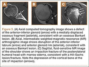
- Associated bony lesions
The incorporation of these imaging advancements into clinical practice supports a more informed approach to managing athletic shoulder injuries, ultimately enhancing recovery prospects and long-term joint function. As diagnostic accuracy improves, the disparity between initial assessment and treatment outcomes shrinks, promising a brighter future for athletes at all levels.
Understanding the Impact of Athletic Shoulder Injuries on Performance and Recovery
Athletic shoulder injuries, particularly those involving the labroligamentous structures, pose a significant threat to an athlete’s performance. Understanding the nuances of these injuries is crucial for both athletes and healthcare professionals. When a labrum or ligament sustains damage, it can compromise shoulder stability and function, leading to diminished performance levels and an increased risk of re-injury. Key factors that affect performance include:
- Range of motion – Limited mobility can hinder an athlete’s ability to execute their sport-specific skills.
- Strength deficits – Injuries often result in weakened muscles, impacting an athlete’s power and endurance.
- Pain management – Chronic pain due to injuries can alter an athlete’s focus and mental resilience, crucial for competitive sports.
Recovery from these injuries requires a tailored approach, as not all athletes will heal at the same rate. MR imaging has emerged as an essential tool for diagnosing these complex conditions, allowing for a more visual representation of the affected areas. The focus of rehabilitation should include:
- Comprehensive assessment – Identifying the exact nature of the injury aids in formulating an effective rehab plan.
- Targeted therapy – Incorporating specific exercises to strengthen the shoulder’s supporting muscles can expedite recovery.
- Gradual return to sport – A step-wise progression can help athletes regain their confidence and functional abilities without risking setbacks.
| Injury Type | MR Imaging Findings |
|---|---|
| Labral Tear | High signal intensity on fluid-sensitive sequences |
| Rotator Cuff Tear | Muscle atrophy and retraction |
| Shoulder Instability | Bone marrow edema and ligamentous injury |
Recommended Protocols for Accurate Diagnosis and Management of Shoulder Labral Tears
- Utilize Advanced Imaging: Implement high-resolution MRI techniques, including MR arthrography, to enhance visualization of the labrum and associated soft tissue structures.
- Conduct Comprehensive Physical Assessments: Perform specific shoulder maneuvers to assess range of motion, strength, and stability, aiding in the correlation of clinical findings with imaging results.
- Integrate Patient History: Consider the athlete’s history, including the type and level of sport played, which can influence both the occurrence and presentation of labral injuries.
Management protocols should be tailored to each individual athlete, balancing surgical interventions with conservative care. Guidelines recommend:
- Personalized Rehabilitation Programs: Develop targeted physical therapy regimens focused on restoring strength and flexibility while avoiding aggravation of the injury.
- Regular Follow-ups: Schedule periodic evaluations to monitor recovery progress and adjust treatment plans as necessary.
- Patient Education: Empower athletes with knowledge about their condition, informing them about the risks and benefits of various treatment options, including potential surgery.
The Conclusion
As the landscape of sports medicine continually evolves, understanding the complexities of labroligamentous injuries in the athletic shoulder is becoming increasingly critical. The insights provided in the latest RSNA Journals article, “Beyond the Cuff: MR Imaging of Labroligamentous Injuries in the Athletic Shoulder,” highlight the pivotal role of advanced imaging techniques in diagnosing and managing these often-overlooked conditions. With an emphasis on the need for comprehensive assessment and awareness, this research provides valuable guidance for clinicians aiming to optimize care for athletes encountering shoulder instability.
As we move forward, staying informed about these developments will enable healthcare professionals to enhance their approach to treatment, paving the way for quicker recoveries and improved outcomes for athletes in all fields. The intersection of imaging technology and clinical practice holds promise for a future where athletes can return to their game stronger and more resilient than ever. Stay tuned for more advancements in this vital area of sports medicine, as we continue to uncover the nuances of athletic injuries and the innovative strategies designed to combat them.


